
Quantifying Tendon Health: An Interview with Prof. Seth O’Neill
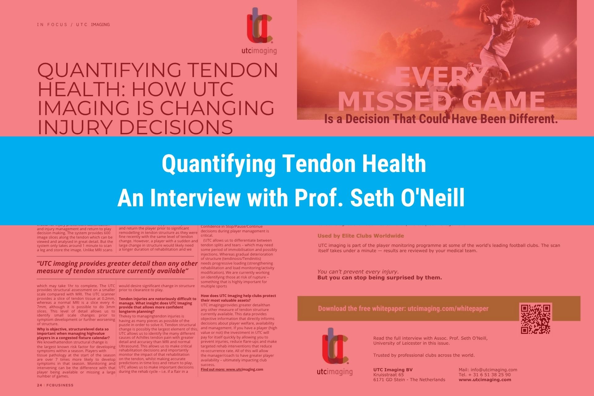
Quicker than MRI and more detailed than conventional ultrasound, UTC scans produce hundreds of ultrafine slices to objectively track tendon collagen, helping clubs identify risk early, steer rehabilitation, and make confident return-to-play decisions. Associate Professor Seth O’Neill of the University of Leicester explains.
What makes UTC Imaging fundamentally different
“It’s the only system in the world that allows you to truly quantify tendon structure (collagen) and monitor change,” says O’Neill. “This is crucial for injury prevention, identification, injury management, and return-to-play decision making.”
The system provides 600 image slices along the tendon, which can be viewed and analysed in great detail. The scan itself takes around one minute to scan a leg and store the image, unlike MRI scans, which may take an hour. The UTC provides structural assessment on a smaller scale than MRI: the UTC scanner captures a slice of tendon tissue at 0.2mm, whereas a normal MRI slice is every 4 to 7mm, although it is possible to do 3mm slices. This level of detail allows clinicians to identify small-scale changes prior to symptom development or further worsening of structure.
Why structure-level data matters in a congested calendar
“We know that tendon structural change is the largest known risk factor for developing symptoms within a season,” O’Neill explains. “Players with tissue pathology at the start of the season are over seven times more likely to develop symptoms in that season. Monitoring and intervening can be the difference between that player being available or missing a large number of games.”
If a player does develop symptoms, the previous scan informs clinicians whether there has been a significant injury or whether the problem has been more of a gradual deterioration. “This can really guide us with return-to-play decisions,” says O’Neill. “A gradual change in structure and sudden flare in tendon pain would mean we are happy to settle pain and return the player prior to significant remodelling in tendon structure, as they were fine recently with the same level of tendon change. However, a player with a sudden and large change in structure would likely need a longer duration of rehabilitation, and we would desire significant change in structure prior to clearance to play.”
Managing notoriously difficult injuries
“The key to managing tendon injuries is having as many pieces as possible of the puzzle in order to solve it,” O’Neill says. “Tendon structural change is possibly the largest element of this. UTC allows us to identify the many different causes of Achilles tendon pain with greater detail and accuracy than MRI and normal ultrasound.”
This allows clinicians to make critical rehabilitation decisions and, importantly, to monitor the impact of that rehabilitation on the tendon, while making accurate predictions on time loss and return to play. “UTC allows us to make important decisions during the rehab cycle,” O’Neill explains. “If a flare in a player’s symptoms occurs and we can clearly see that the tendon structure is improving, we can make a decision to continue rehab or pause at the same level. Whereas if that flare in symptoms occurs with a deterioration in structure, we can make the important decision to stop rehab or backtrack and regress. Confidence in stop, pause, or continue decisions during player management is critical.”
UTC also allows clinicians to differentiate between tendon splits and tears, which may need a period of immobilisation and possibly injections, and gradual deterioration of structure such as tendinosis or tendinitis, which needs progressive loading, strengthening rehabilitation, and load monitoring or activity modification. O’Neill’s team is currently working on identifying those at risk of rupture, something highly important across multiple sports.
Protecting a club’s most valuable assets
“UTC imaging provides greater detail than any other measure of tendon structure currently available,” says O’Neill. “This data provides objective information that directly informs decisions about player welfare, availability, and management.”
Whether the player in question carries a high transfer value or not, the investment in UTC pays for itself quickly by allowing clubs to prevent injuries, reduce flare-ups, and make targeted rehab interventions that reduce re-occurrence rate. All of this gives the manager and coaching staff greater player availability, ultimately impacting club success.
Source: interview with Assoc. Prof. Seth O’Neill (University of Leicester), published in FCbusiness, issue 173, “In Focus: UTC Imaging” (p. 24).