Epic Equine Comeback After UTC-Guided Tendon Rehab
Lady Ladegaard, a Danish Warmblood mare born in 2015, presented with a history of superficial digital flexor tendon (SDFT) injuries. Her case highlights the critical role of UTC imaging in managing tendon injuries in equine athletes.
Initial Presentation
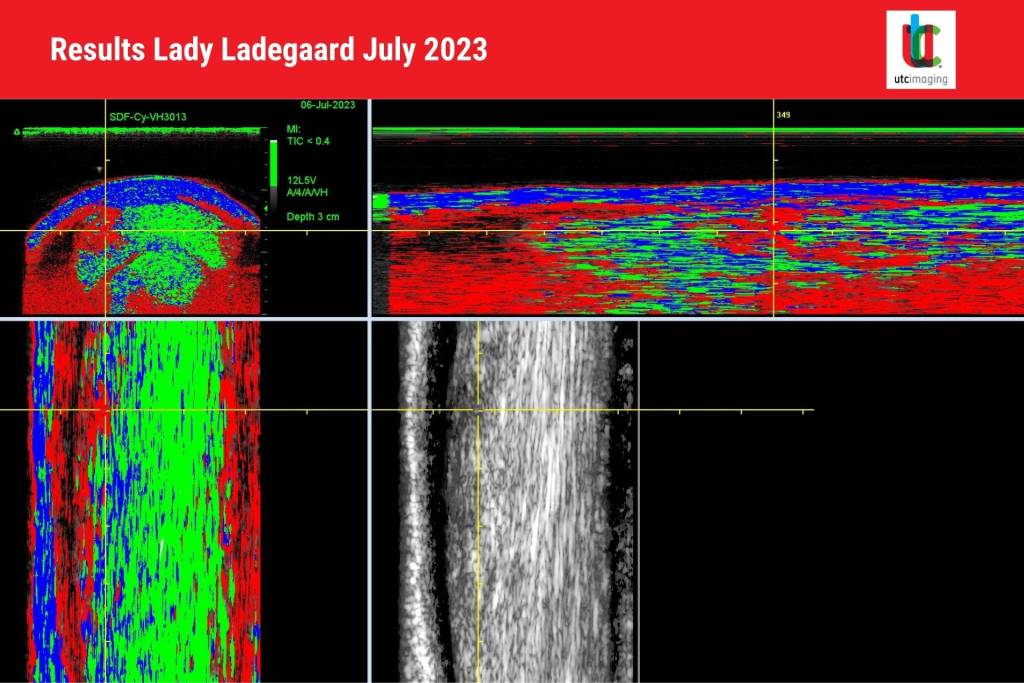
In July 2023, Lady Ladegaard was brought to Dr. Johan Lenz of the Lenz Hastfokus clinic, with acute swelling in her left front SDFT. The injury was confirmed as tendinitis via ultrasound and UTC imaging. UTC revealed fibrosis and scarring in the tendon, as well as a hypoechoic lesion indicating a new rupture. The mare began a structured rehabilitation program, involving controlled exercise and medication. She was confined to a small paddock and underwent daily walking sessions to minimize strain.
Progress and Monitoring
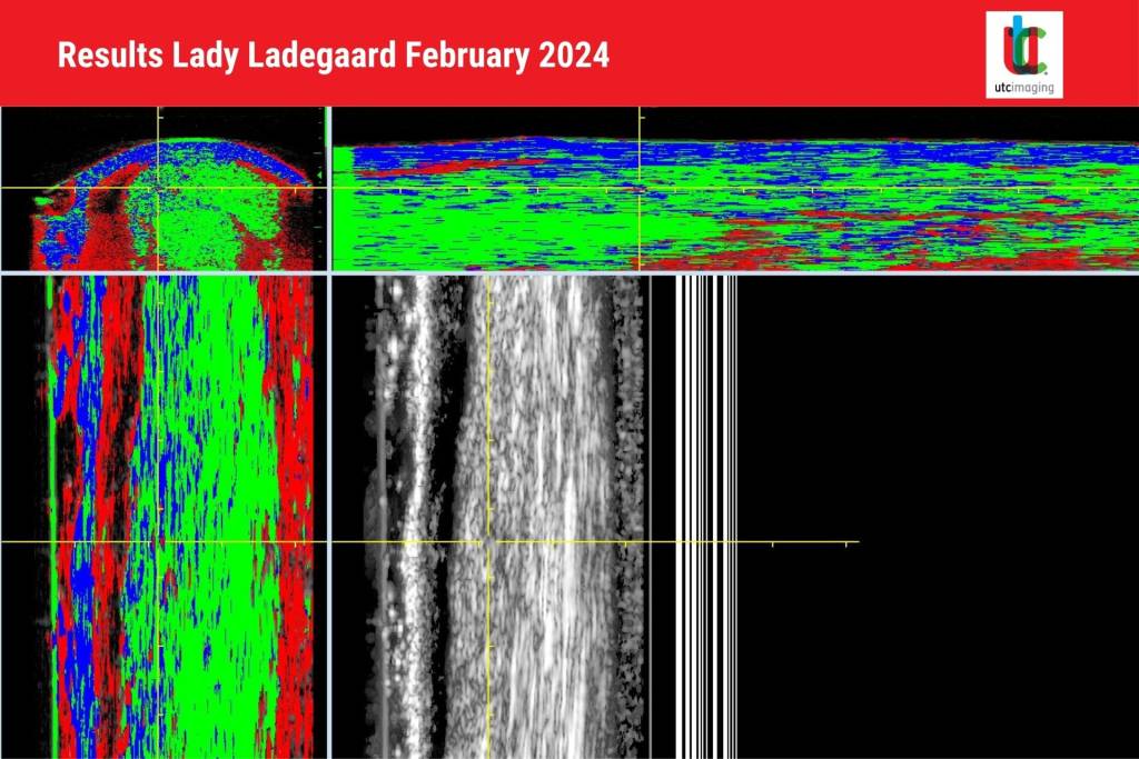
After eight months of rest, a follow-up in February 2024 showed significant healing. The UTC scan demonstrated reduced Type 3 fibers (indicative of poor tendon structure) and increased collagen organization, marking good tissue regeneration. The rehabilitation program progressed to light riding.
Two months later, in April 2024, another UTC scan confirmed acceptable adaptation of the scar tissue under increased workload. Controlled trotting and galloping were gradually introduced.
Return to Competition
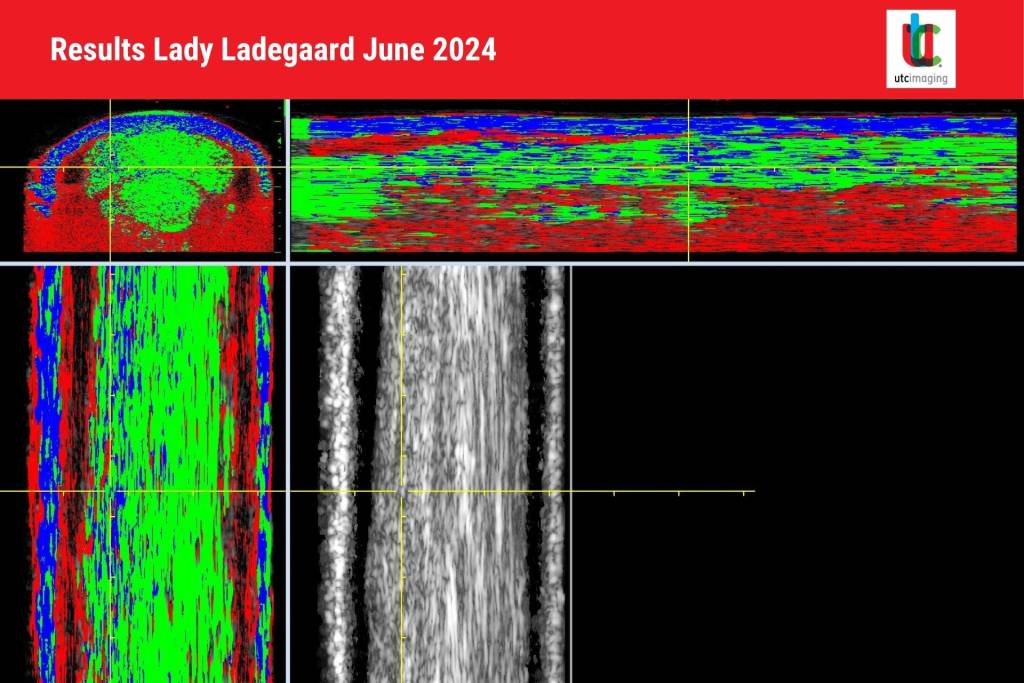
By June 2024, Lady Ladegaard had returned to competition, achieving excellent results in the L:A level. The UTC scan showed improved tissue integrity with a notable increase in Type 1 fibers, reflecting robust tendon structure. She was cleared for higher-intensity training, aiming to compete at the MSV level in the fall.
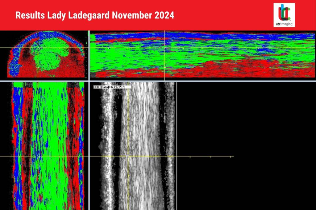
Final Assessment
In November 2024, Lady Ladegaard was in near-full training. The UTC imaging confirmed minimal remaining abnormalities, with well-organized tendon fibers. She was allowed to continue training at full capacity, with a final follow-up recommended within three months.
The Role of UTC in Equine Sports Medicine
UTC imaging proved indispensable in this case. Unlike conventional ultrasound, UTC offers three-dimensional visualization and precise tissue characterization. It distinguishes between four echo types:
- Type 1 (green): Intact, aligned fibers.
- Type 2 (blue): Wavy or less aligned fibers.
- Type 3 (red): Small fibrillar components indicating disorganization.
- Type 4 (black): Amorphous tissue and fluid.
This technology enabled Dr. Lens to monitor subtle changes in Lady Ladegaard’s tendon structure throughout her recovery. Early detection of degenerative changes ensured targeted therapy, while successive scans informed adjustments to her rehabilitation plan.
Key Benefits of UTC:
- Accurate Diagnosis: Differentiates between acute and chronic injuries.
- Guided Rehabilitation: Tracks progress and adjusts exercise intensity based on tissue condition.
- Prognostication: Predicts outcomes by monitoring repair quality and risk of re-injury.
- Prevention: Identifies subclinical degradation, reducing the likelihood of future injuries.
Conclusion
Lady Ladegaard’s recovery illustrates the potential of UTC imaging to transform equine sports medicine. By offering detailed insights into tendon health, this innovative technology enables veterinarians to tailor treatments and guide rehabilitation with unparalleled precision. For equine athletes, UTC represents a pathway to faster recovery, reduced recurrence, and a prolonged competitive career.