UTC Case Study Show jumper: insights in Equine Tendon Care:

During the Veterinary Sport Horse Congress in Amsterdam, a focused symposium on UTC imaging gathered a dedicated group of practitioners, with Frans van Toor from SMDC in Heesch, the Netherlands, at the helm. Among the speakers, Dr. Van Schie offered a compelling session on UTC, with presenting an UTC Case Study on a show jumper.
This session emphasized the critical need for a nuanced understanding of Ultrasound Tissue Characterization. Dr. Van Schie’s presentation, titled “Understanding Tendon Injuries: The Clinical Challenge,” served as a foundation for exploring the transformative impact of UTC imaging in diagnosing and treating tendon injuries in equine athletes.
As we unpack the details of this presentation, we spotlight the “UTC Case Study on Show jumper,” highlighting the synergy between theoretical insights and their practical implications in the realm of equine tendon diagnostics.
The Complexity of Tendon Pathologies
Tendon injuries present a unique challenge in clinical practice due to their diverse pathology. Often, what appears as a recent acute injury may, in fact, have a prolonged history, characterized by gradual onset and underlying degeneration that initially shows no clinical signs. Such degenerative changes forecast a gloomier prognosis, with slower recovery and a tendency for relapse. This diversity necessitates a comprehensive diagnostic approach, as there is no universal “one size fits all” treatment strategy.
The Multistage Nature of Tendon Damage
It’s crucial to discern the stages of injury within the tendon tissue. A seemingly recent injury could overlay chronic degenerative changes, necessitating deeper investigation. This realization led to the development of advanced tissue characterization techniques to distinguish between fresh injuries and long-standing alterations.
The Genesis of Tissue Characterization
The journey of tissue characterization began in 1979 with Van Schie’s acquisition of his first ultrasound device. Initially content with observing the disappearance of the ‘black hole’ in the scans, he later realized the need to understand what these changes represented. By examining the tendon after sacrificing the horse, a strong skepticism emerged about the correlation between conventional grayscale imaging and tendon integrity. Recognizing the inherent limitations of two-dimensional imaging for a three-dimensional structure, the move towards a more scientific approach was clear.
From Art to Science: The Development of UTC Imaging
Traditional ultrasound was often seen as more art than science due to its reliance on the operator’s skill for image acquisition and interpretation. To address this, a method to compile images was devised. The UTC tracker was developed to standardize the imaging process, moving the transducer along the tendon and capturing transverse images every 0.2 millimeters. This automation provides consistent, reproducible results with dedicated presets for different tendons, focusing not on the aesthetics of the image but on capturing raw, real radio-frequency signals.
Operator-Independent Imaging
With UTC, the imaging process becomes operator-independent, ensuring reliability and repeatability. The system’s presets allow for scanning various tendons with standardized settings, ensuring that images reflect true tissue characteristics rather than operator preference. This technology provides a three-dimensional data block that can be scrolled through in three different planes simultaneously.
Sensitivity to Load Effects and Real-Time Imaging
The precision of the UTC tracker and the sensitivity of the UTC algorithms allow for the detection of subtle load effects in the tendon. With its ability to move the transducer along the tendon and collect images in precise increments, it offers a unique view of the tendon’s condition. This capacity results in transverse, sagittal, and coronal images that are stable, reliable, and allow detailed information on the tendon tissue.
Ground Truth Database and Tendon Classification
Establishing a Reference Standard
The development of UTC was validated on a comprehensive database of 148 tendons, thoroughly examined to establish a ground truth. This database included detailed UTC imaging slices correlated with histological, biochemical, and mechanical loading tests data.
Echo Type Classification
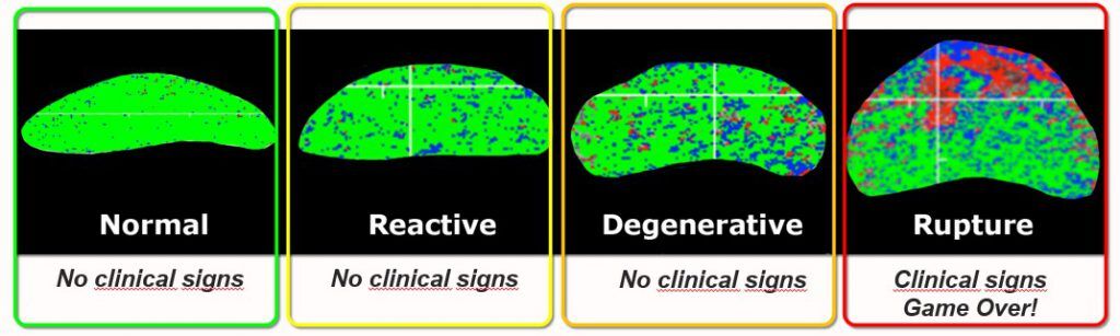
Through this robust analysis, tendons were classified into four echo types, signifying different tissue characteristics:
Type I (green): Intact and aligned tendon fascicles
Type II (blue): Discontinuous or wavy tendon fascicles above the spatial resolution
Type III (red): Mainly fibrillar components
Type IV (black): Amorphous cellular components and fluid
This classification system, derived from the ground truth database, ensures excellent reliability and sensitivity, surpassing 90% efficacy.
Superior Reliability in Tendon Imaging
In every instance, UTC has demonstrated remarkable reliability and sensitivity, exceeding 90%. This level of precision is substantially greater when compared to traditional ultrasound methods.
The Dual Purpose of UTC: Detection and Monitoring
At present, UTC is employed chiefly for two objectives: monitoring load effects on tendons and facilitating early detection of lesions. The goal is to intervene before it’s too late and distinguishing whether a tendon injury is a recent, isolated event or a recent development superimposed on chronic alterations. The insights gained from UTC staging are instrumental in devising targeted therapeutic strategies and refining rehabilitation protocols.
UTC Case Study: Monitoring an Olympic Show jumper
Initial Observations and Progressive Changes
Consider the case of an Olympic show jumper from the Dutch team, first scanned in 2011. Initial scans revealed a flawless superficial digital flexor tendon. By February 2012, subtle changes began to manifest—represented by an increase in blue on the scan, indicating minor swelling, yet without clinical symptoms. By April, the tendon’s architecture had altered significantly, a development that, while not clinically evident, was concerning. Neither rider nor trainer was willing to modify the training schedule.
Alarming Continuum of Pathology
As the months progressed, the situation deteriorated alarmingly. By early June, what had begun as benign had escalated to a severe injury with both red and black areas on the UTC scan, signaling extensive damage and the presence of fluid or cellular components.
Interpreting UTC Data for Targeted Action
When analyzing the UTC images, it’s clear that the injury to the superficial flexor tendon was severe, with extensive areas indicated by red and black on the scan. Despite the acute stage of the injury, scar tissue at the center was indicative of the underlying degenerative issue. Unfortunately, in this case, the horse was ultimately sacrificed.
Case Study: Managing a Racehorse’s Tendon Injury
The Challenge of a Chronic Injury
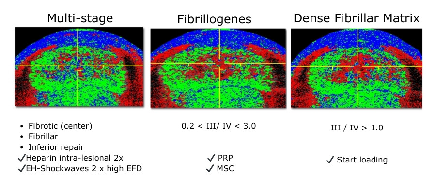
In a detailed case study of a racehorse, UTC played a pivotal role in managing a complex tendon injury featuring a central chronic scar, likened to a “stone in the shoe.” Initially, the horse displayed a mix of acute and chronic injury stages within the tendon.
Intervention Strategies
The management strategy included targeted injections and heavy shockwave therapy aimed at removing the chronic scar and improving perfusion. Once this ‘stone’ was removed, the treatment shifted to Platelet-Rich Plasma (PRP) and stem cell therapies, guided by the relative ratio of red to black on the UTC scan, which represents the density of the fibrillar matrix.
Monitoring and Adjusting Treatment
As the healing process was monitored, the increase in red and the emergence of green and blue indicated a progressing structure, prompting the initiation of loading exercises. This early intervention and structured rehabilitation, informed by UTC staging, allowed for a quicker return to training activities than typically expected.
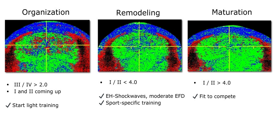
The Outcome
Over the course of six to seven months, the horse underwent a successful recovery, transitioning from initial treatments to sport-specific training. The case exemplifies how UTC can guide rehabilitation, providing a window into the healing stages of tendon injuries.
Conclusion: Advancing Tendon Care with UTC
In this UTC Case Study on show jumpers, we can conclude that the essence of effective tendon management lies in early detection and precise staging of injuries. As demonstrated by the case of the Olympic show jumper, UTC is crucial for early intervention and preventing clinical symptoms.
Staging the injury accurately—distinguishing between single and multiple stages—enables targeted therapy that’s tailored to the individual’s condition. While not all tendons may fully return to their pre-injury state, the aim is to minimize the risk of relapse through careful monitoring and guided rehabilitation using UTC insights.
In summary, UTC is an invaluable tool in our arsenal, shaping a proactive approach to tendon health that prioritizes early detection and informed treatment strategies for better outcomes and sustained athletic performance.
Enhancing Clinical Outcomes
The introduction of Ultrasound Tissue Characterization (UTC) represents a significant advancement in the diagnosis and treatment of tendon injuries. By moving from a subjective art form to a standardized, scientific approach, clinicians are now equipped with a powerful tool to assess tendon integrity and guide treatment, ultimately enhancing patient outcomes.